 FiberRemedy
Stain Classification & Redox Chemistry
Protein stains: temperature and first treatment
A fresh food or blood stain is easier to assess before a hot wash or dryer cycle changes it.
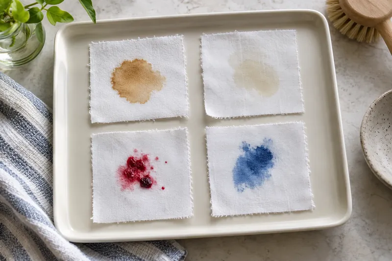
Before you start
A fresh food or blood stain is easier to assess before a hot wash or dryer cycle changes it. Use the care label to establish whether rinsing is permitted, then choose a fabric-compatible stain product. Older mixed stains may need more than one permitted treatment, but each should have a clear purpose.
For almost every household cleaning problem—greasy dishes, muddy floors, waxy collar rings—hotter water means faster cleaning. That intuition fails catastrophically the moment a drop of blood, raw egg, milk, or meat juice lands on a cotton bedsheet or dress shirt. Running a fresh bloodstain under steaming 55 °C (131 °F) tap water turns a water-soluble red liquid into a permanently bonded, rust-brown rubbery patch in less than ten seconds. Understanding globular protein thermodynamics (ΔG_unfold), cysteine disulfide interchange (-SH → -S-S-), and heme iron oxidation (Fe²⁺ → Fe³⁺) explains why protein stains obey the strict Cold Water + Saline + Protease Rule. Cross-check all six household protein stain protocols in our Stain Chemistry × Textile Fiber Safety Lookup Matrix.
Native Globular Structure vs. Thermal Denaturation (T_m ≈ 42–68 °C)
Whole human blood contains roughly 150 g/L of hemoglobin inside red blood cells (erythrocytes), alongside 70 g/L of plasma proteins (serum albumin, immunoglobulins, and fibrinogen, which polymerizes into insoluble fibrin blood clots via the enzyme thrombin). Similarly, egg white is a 10% aqueous solution of ovalbumin, ovomucoid, and conalbumin.
At human body temperature (37 °C / 98.6 °F) and below, these proteins exist in their folded native tertiary conformation:
- Hydrophilic Exterior Shell: Polar and charged amino acid side chains (
lysine,arginine,glutamate,aspartate) point outward into the surrounding water, forming hydration shells that make the folded protein sphere water-soluble. - Buried Hydrophobic Core: Non-polar hydrocarbon side chains (
leucine,isoleucine,valine,phenylalanine,tryptophan) and reactive free cysteine thiol groups (-SH) are tucked tightly inside the core, hidden from the fabric fibers.
When you pour hot water (> 42–50 °C / 108–122 °F) onto the fabric, thermal kinetic energy (k_B · T) overwhelms the weak hydrogen bonds (~12–20 kJ/mol) holding that tertiary fold together:
Three-Step Thermal Coagulation of Protein Stains on Textile Fibers (T > 42–55°C):
  1. Reversible Unfolding (Denaturation):
     Native Globular Protein (Water-Soluble, Hydrophobic Core Buried)
       ──[Heat ≥ T_m]──>  Unfolded Polypeptide Chain (Hydrophobic Residues & -SH Exposed!)

  2. Physical Entanglement & Intermolecular Beta-Sheet Aggregation:
     Exposed hydrophobic side chains flee the hot water by wrapping tightly around cotton/polyester
     microfibrils and locking with neighboring unfolded chains into insoluble β-sheet aggregates.

  3. Covalent Disulfide Cross-Linking (Thiol-Disulfide Interchange):
     2 Protein-SH + ½ O₂  ──>  Protein-S–S-Protein (Covalent Disulfide Bridge, ~250 kJ/mol!) + H₂O
Once unfolded polypeptide chains wrap around the microscopic lumen and crenulations of a cotton fiber and cross-link via covalent disulfide bridges (-S-S-), they are no longer water-soluble monomers—they have cured into a solid elastomeric glue (identical to how transparent liquid egg white turns into opaque solid rubber in a frying pan).
| Protein Stain Component | Source Soil | Thermal Denaturation Onset (T_onset) |
Midpoint Coagulation Temp (T_m) |
Safe Wash Temperature Ceiling |
|---|---|---|---|---|
| Fibrinogen / Fibrin | Whole blood (clotting matrix) | 42–45 °C (108–113 °F) |
~55 °C (131 °F) |
≤ 28 °C (82 °F, Cold) |
| Conalbumin (Ovotransferrin) | Raw egg white | 53–56 °C (127–133 °F) |
~61 °C (142 °F) |
≤ 30 °C (86 °F, Cold) |
Hemoglobin (HbA) + Heme Fe²⁺ |
Red blood cells | 54–58 °C (129–136 °F) |
~64 °C (147 °F) |
≤ 28 °C (82 °F, Cold) |
Serum Albumin (HSA / BSA) |
Blood plasma, meat juice | 55–58 °C (131–136 °F) |
~62 °C (144 °F) |
≤ 30 °C (86 °F, Cold) |
| Myoglobin | Beef, pork & poultry purge | 55–60 °C (131–140 °F) |
~65 °C (149 °F) |
≤ 30 °C (86 °F, Cold) |
| Ovalbumin | Egg white (54% of protein) | 70–74 °C (158–165 °F) |
~78 °C (172 °F) |
≤ 30 °C (86 °F, Cold) |
Why Isotonic Saline (1% NaCl) Beats Plain Water on Bloodstains
When treating fresh or semi-dried blood on delicate fabrics (especially wool, cashmere, or silk where enzyme detergents are forbidden), dissolving 1 teaspoon (~6 g) of ordinary table salt (NaCl) per 2 cups (500 mL) of cold water (~1.0–1.2% w/v, ionic strength I ≈ 0.17–0.20 M) dramatically outperforms plain tap water for two biophysical reasons:
- The Debye-Hückel “Salting-In” Effect on Globulins: Blood plasma contains a high fraction of globulins (
immunoglobulinsandfibrinogen), which by definition are insoluble or poorly soluble in pure deionized/low-ion water (I → 0) because oppositely charged patches on adjacent globulin molecules attract each other electrostatically and precipitate. Adding0.15–0.20 MofNa⁺andCl⁻ions creates an ionic atmosphere (Debye screening lengthκ⁻¹ ≈ 0.7 nm) around those charged surface patches, keeping globulins dissolved so they flush out of the yarn! - Preventing Erythrocyte Membrane Ghosts From Trapping in Yarn Pores: In pure tap water (
hypotonic), intact red blood cells swell osmotically and burst (hemolysis), plastering sticky lipid cell-membrane fragments onto hydrophobic fibers while releasing free hemoglobin. Cold0.9%–1.0%isotonic saline suspends intact cells and solubilizes plasma clots cleanly.
Hemoglobin Aging Chemistry: Oxyhemoglobin → Methemoglobin → Hemichrome
Why does an untreated bloodstain turn from bright crimson to dark brown-orange over 48 hours and become ten times harder to remove? Each hemoglobin tetramer carries four iron-porphyrin heme groups bound to the protein via a proximal histidine residue:
- Fresh Crimson Blood (
Oxyhemoglobin,Fe²⁺-O₂): Ferrous iron (Fe²⁺) is reversibly coordinated to dioxygen; protein globin chains are still native and water-soluble. - Aged Brown Blood (
Methemoglobin,Fe³⁺→Hemichrome): Atmospheric autoxidation converts ferrous iron (Fe²⁺) into ferric iron (Fe³⁺, methemoglobin) while releasing superoxide radicals (O₂•⁻). Over hours to days, the distal histidine side chain coordinates directly to theFe³⁺center (bis-histidyl hemichrome), cross-linking the denatured globin protein and leaving an insoluble ferric oxide/porphyrin rust-brown core embedded in the fiber.
Step-by-Step Protocol: Fresh vs. Dried/Heat-Set Blood & Protein Stains
Protocol A: Cotton, Linen & Synthetics
- Immediate Cold Reverse Flush (
15–25 °C): Hold the stained area inside-out under a forceful stream of cold tap water so hydrostatic pressure pushes blood clots out the face of the fabric rather than driving them deeper through the weave. - Cold Saline + Subtilisin Protease Soak (
30–60 Minutes): Submerge in1 quartof cold water containing2 teaspoonsof non-iodized salt (NaCl) plus1 tablespoonof liquid enzyme detergent containing subtilisin protease (Laundry Enzymes Explained). Protease snips coagulated fibrin and globin chains into short water-soluble peptides. - Oxidize the Residual Heme Ring (
Only After Protein Is Gone): If a faint tan/orange heme shadow remains on white cotton, apply a few drops of3%hydrogen peroxide (H₂O₂) (which fizzes via blood catalase enzyme:2H₂O₂ → 2H₂O + O₂↑) or soak in warm sodium percarbonate (Sodium Percarbonate vs. Hypochlorite Guide). (If an old bloodstain has left an inorganic ferric ironFe³⁺rust spot that ignores peroxide, chelate the iron with a 5-minute dab of2%oxalic acid / Bar Keepers Friend, then rinse!)
Protocol B: Silk, Merino Wool & Cashmere (Zero Protease, Zero Percarbonate!)
Because silk fibroin and wool keratin are proteins, never use protease detergent or alkaline pH 10.5 OxiClean. Soak the garment in cold 1.0% NaCl saline (2 tsp salt per quart cold water) for 30 minutes, gently tamp with a pH 6.5–7.0 non-enzymatic wool/silk wash, and—if a heme shadow remains on light fabric—dab with cool 3% H₂O₂ for 5 minutes and rinse cold.
Practical check: what to observe
Avoid rubbing a fragile textile hard enough to distort it. After washing, inspect the area before tumble drying. If the item has biological contamination that needs disinfection, follow relevant product and public-health instructions rather than treating appearance as proof of hygiene.
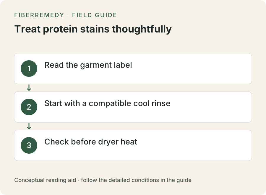
- Read the garment label
- Start with a compatible cool rinse
- Check before dryer heat
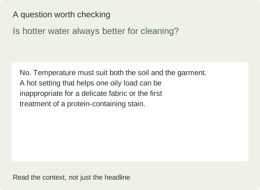
Is hotter water always better for cleaning?
No. Temperature must suit both the soil and the garment. A hot setting that helps one oily load can be inappropriate for a delicate fabric or the first treatment of a protein-containing stain.
For more context, see the topic FAQ and glossary. A reference value or example should be read with its units, assumptions and product-specific conditions.
Sources and scope
The references below were supplied with the original manuscript. A reference is not evidence that every numerical claim has been independently checked. See the source library and our verification status.
- Biochemistry (Voet & Voet) – Thermal Denaturation, Hydrophobic Collapse, and Disulfide Cross-Linking of Globular Proteins
- Forensic Science International – Hemoglobin Oxidation States (Oxyhemoglobin → Methemoglobin → Hemichrome) and Bloodstain Persistence on Textiles
- Journal of Surfactants and Detergents – Removal Kinetics of Thermally Coagulated Blood and Ovalbumin from Cotton Fabrics
Put the explanation to work
Continue reading
Spot a questionable claim or a calculation issue? Read our correction process and contact information. Examples and illustrations are educational; they do not establish product suitability or a laboratory result.